Question 24
(Multiple Choice)
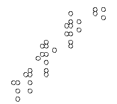
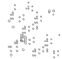
In an unfolded (random coil) protein, amino acid residues are exposed to the solvent and share more or less the same environment, whereas each residue in a folded protein has its own unique neighborhood. This fact can be exploited in using NMR to study protein folding. The two schematic diagrams below represent two-dimensional NMR spectra for the same protein in either its folded (native) or unfolded state. The chemical shifts, which depend on the local neighborhood of each atom, are plotted in these diagrams. Which diagram (A or B) do you think corresponds to the folded protein?
A)
B)
Answer